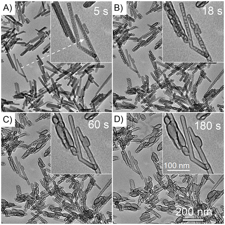
Figure 1. TEM images of Na-dawsonite nanorods at (A) 5, (B) 18, (C) 60, and (D) 180 s exposure under 120 kV-electron-beam irradiation.

The insets are enlarged TEM image of the area pointed by the arrow in (A).

The insets are enlarged TEM image of the area pointed by the arrow in (A).