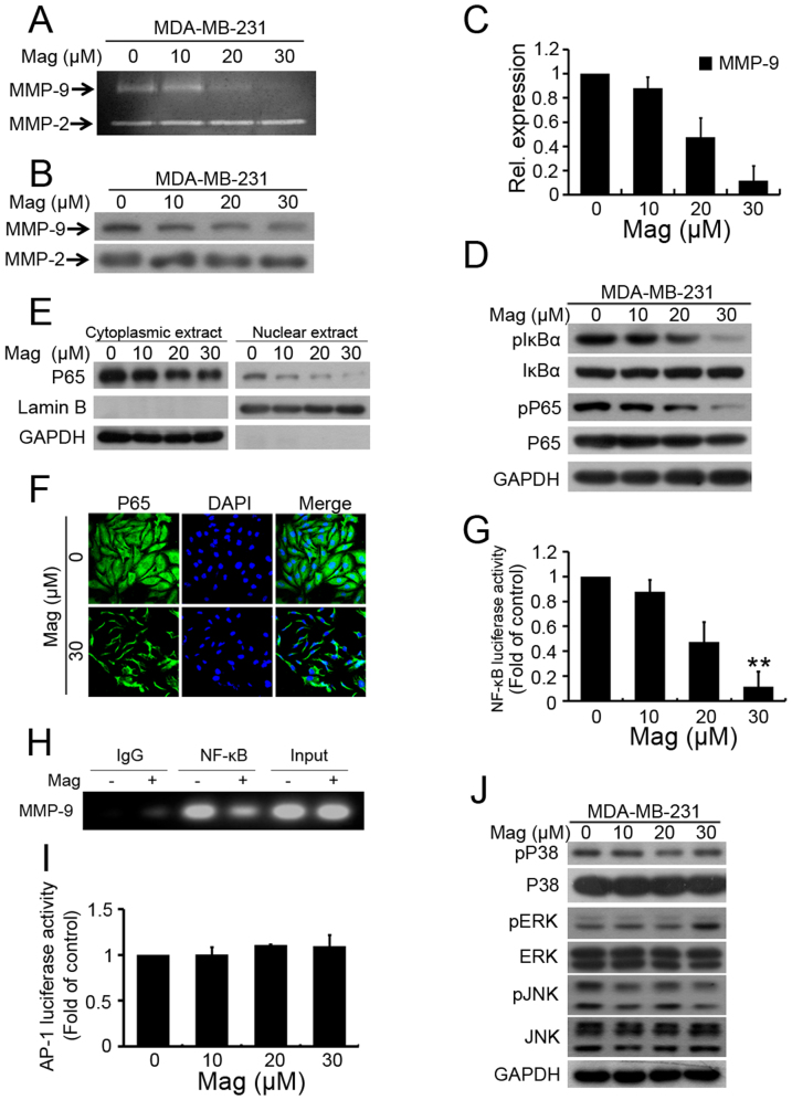
Figure 3. Mag suppresses breast cancer cell invasion through the inhibition of MMP-9 via NF-κB pathway.
MDA-MB-231 cells were treated with increasing concentrations of Mag for 24 h. (A): The activity of MMP-9 was assessed using the concentrated conditioned medium (CM) by gelatin zymography. The concentration of CM protein was measured by Bradford assay. (B): Western blot was performed using antibodies indicated from CM of the Mag treated cells. The concentration of CM protein was measured by Bradford assay. (C): MMP-9 gene expression was detected by Real-time PCR analysis. GAPDH was used here as a housekeeping gene. (D): MDA-MB-231 cells were treated with increasing concentrations of Mag for 24 h, Western blot was performed using antibodies indicated. (E): Cytoplasmic and nuclear fractions of MDA-MB-231 cells were isolated, the concentration of nuclear and cytoplasmic protein was measured by Bradford assay, the same amount of nuclear and cytoplasmic protein was subjected to SDS gel, and Western blot were performed with anti-P65, GAPDH and Lamin B antibodies. (F): MDA-MB-231 cells were treated with Mag at 0 and 30 μM for 24 h. For immunofluorescence analysis, cells was stained with an anti-P65 antibody and DAPI and observed by confocal microscopy. (G): MDA-MB-231 cells were transfected with the NF-κB luciferase reporter construct for 4 h and then treated with Mag. Luciferase activity was measured using the Luciferase assay system. (H): MDA-MB-231 cells treated with Mag (20 μM) were processed for ChIP assay. Immunoprecipitation was performed with NF-κB p65 or IgG as a control. The NF-κB binding site of MMP-9 promoter was detected by PCR. (I): MDA-MB-231 cells were transfected with the AP-1 luciferase reporter construct for 4 h and then treated with Mag. Luciferase activity was measured using the Luciferase assay system. (J): Western blot was performed using antibodies indicated. *, P < 0.01, **, P < 0.001 vs 0 μM. (I): Real-time PCR analysis MMP-9 expression lysates of tumor samples. **, P < 0.001 vs vehicle. (J): Western blot analysis of lysates of tumor samples using indicated antibodies. Full-length blots/gels are presented in Supplementary Figure 1 and 2.

