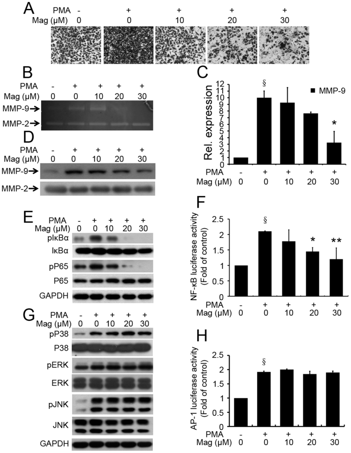
Figure 4. Mag overcomes the facilitative effects of PMA on invasion of MDA-MB-231 cells.
(A): MDA-MB-231 cells were preincubated with Mag for 30 min, and then cell suspension containing Mag and PMA (80 nM) was seeded onto the upper chamber wells. After incubation for 24 h at 37°C, the filter was fixed and stained with 0.2% crystal violet powder (15 min). Then randomly chosen fields were photographed (×100). Scale bar = 300 μm. (B): MDA-MB-231 cells were pretreated with the indicated concentration of Mag for 30 min and then stimulated with 80 nM PMA for 24 h. The activity of MMP-9 was assessed using the concentrated CM by gelatin zymography. The concentration of CM protein was measured by Bradford assay. (C): MDA-MB-231 cells were pretreated with the indicated concentration of Mag for 30 min and then stimulated with 80 nM PMA for 24 h. MMP-9 gene expression was detected by Real-time PCR analysis. GAPDH was used here as a housekeeping gene. (D): The protein expression of MMP-9 was assessed using the concentrated CM by Westen blot. The concentration of CM protein was measured by Bradford assay. (E): Western blot was performed using antibodies indicated. (F): MDA-MB-231cells transfected with pNF-κB-luc reporter plasmid for 24 h were pretreated with Mag for 30 min and then exposed to PMA for another 20 h. NF-κB firefly luciferase activity was measured using the Luciferase assay system. § P < 0.05 vs control (without PMA treatment); *P < 0.05, **P < 0.01 vs PMA-only group. (G): Western blot was performed using antibodies indicated. (H): MDA-MB-231cells transfected with pAP-1-luc reporter plasmid for 24 h were pretreated with Mag for 30 min and then exposed to PMA for another 20 h. AP-1 firefly luciferase activity was measured using the Luciferase assay system. § P < 0.05 vs control (without PMA treatment). Full-length blots/gels are presented in Supplementary Figure 3.

