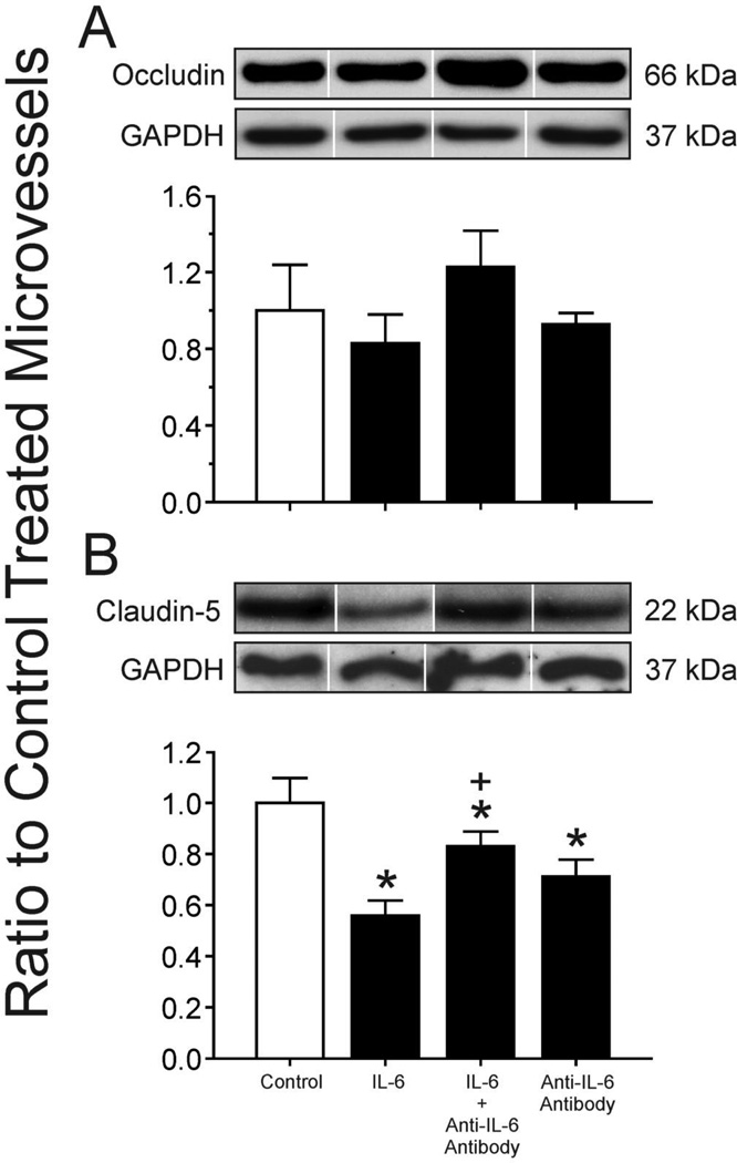
Figure 4.
A Occludin expression in isolated cerebral microvessels from adult sheep after incubation with IL-6 protein (100 ng/ml), IL-6 protein (100 ng/ml) preincubated with IL-6 neutralizing monoclonal antibodies (1 µg/ml) and IL-6 neutralizing monoclonal antibodies (1 µg/ml) alone. Representative Western immunoblots for occludin and GAPDH. Results shown as ratio to PBS control treated microvessels. Open bar is control treated microvessels set to a value of 1; closed bars plotted for IL-6 alone, IL-6 plus anti-IL-6 antibody, and anti-IL-6 antibody alone as indicated on the x-axis. n=5 for each group. Values are mean ± SEM.
B Claudin-5 expression in isolated cerebral microvessels from adult sheep after incubation with IL-6 protein, IL-6 protein preincubated with IL-6 neutralizing monoclonal antibodies and IL-6 neutralizing monoclonal antibodies alone. IL-6 protein and IL-6 neutralizing monoclonal antibody concentrations as for figure 4 A. Representative Western immunoblots for claudin-5 and GAPDH. Results shown as ratio to PBS control treated microvessels. Open bar is control treated microvessels set to a value of 1; closed bars plotted for IL-6 alone, IL-6 plus anti-IL-6 antibody, and anti-IL-6 antibody alone as indicated on the x-axis. n=5 for each group. Values are mean ± SEM. * P< 0.05 vs. control, + P < 0.05 vs. IL-6 alone and anti-IL-6 antibody alone.

