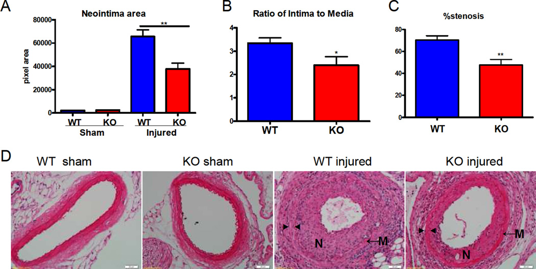
Figure 1. Deletion of mPGES-1 reduced neointimal formation.
Neointimal area (A), the ratio of intima to media (B) and stenosis (C) were reduced significantly in mPGES-1 KO mice compared to WT controls. (*: p<0.05; **: p < 0.01. n=4 per group for sham-operated animals; n=13 for WT injured animals; n=12 for KO injured animals). Representative H&E staining of cross sections from sham operated or wire-injured arteries is shown (D). N denotes neointima; M denotes media; ► indicates internal elastin; ◄ indicates external elastin. Scale bar denotes 20 µm.

