
Figure 4.
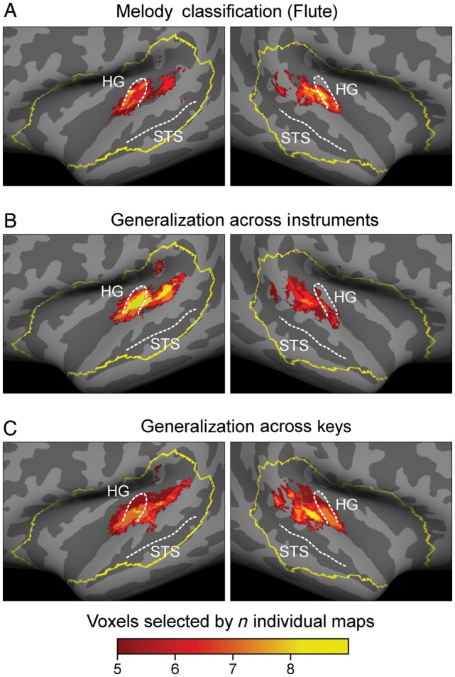
Multiparticipant consistency maps of discriminative voxels shown on a standard brain surface of the temporal lobes. The yellow outline illustrates the anatomically defined region of interest from which all analyses started from. All group maps were created by summation of the individual discriminative voxel maps of all 8 participants. Each map was thresholded such that a voxel had to be selected in at least 5 individuals to appear on the group map. Color coding indicates consistency across participants. Across all classifications no lateralization bias was found (c.f. Supplementary Table S1). For group map of melody classification on piano see Supplemental Figure S1.