Figure 6.
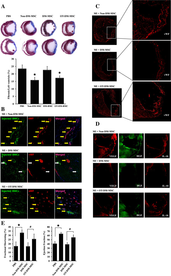
Cardiac fibrosis, expressions of angiogenic factors, and cardiac function were ameliorated by injection of OT-DM-MSC in infarcted myocardium. (A) Representative images showed fibrosis was significantly reduced in Non-DM-MSC and DM-MSC-injected heart. (B) Representative images of GFP-labeled MSCs (green) injected in infarcted myocardium showed expression of von Willebrand factor (vWF, red). The merged images showed vWF-expressing MSC (yellow arrows) and none-expressing MSC (white arrows). (C) Distribution and degree of vWF expression in the whole heart. Less vWF and restored vWF were observed in DM-MSC injected heart and DM-MSC injected heart after MI, respectively. (D) Expressions of vascular endothelial growth factor (VEGF), hepatocyte growth factor (HGF), and interleukin-10 (IL-10) in infarcted myocardium were examined. (E) Echocardiographic evaluations were performed 2 weeks after cell injection. * p < 0.01 vs PBS; # p < 0.01 vs DM-MSC.

