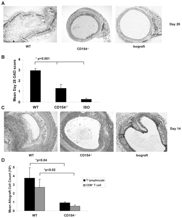
Figure 1. CD154−/− recipient mice have significantly attenuated OAD, with CD8+ T-cell predominance similar to WT airway allografts.
C57BL/6 WT recipient mice or C57BL/6 CD154−/− mice were transplanted with trachea from BALB/c mice and compared to syngeneic isograft recipients of C57BL/6 tracheae. (A) Representative day 28 histopathology from each treatment group; tracheal grafts were fixed, stained with H/E as per materials and methods. (B) Mean OAD scores with bars representing mean values + SEM for treatment group with p-values determined by rank sum test. (C) Representative day 14 histopathology from each treatment group; tracheal grafts were fixed, stained as in (A). (D) Total lymphocyte counts were determined by determined by hemocytometer-based cell count, followed by fluorescent dye-conjugated anti-CD4 and anti-CD8 Ab staining and flow cytometric analysis. Total T lymphocyte count determined as sum of CD4+ and CD8+ fraction. Note that too few cells were recovered from isografts to quantitate T lymphocytes. Bars represent mean values ± SEM of cell counts for treatment group with p-values determined by rank sum test for paired samples and Kruskal–Wallis test for multiple groups. Results represent a minimum of three independent experiments with 8–10 mice per group.

