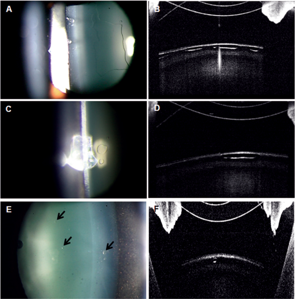
Figure 2.

Anterior segment photographs and optical coherence tomography findings in Case 2. One day after ICL implantation, UDVA was 20/20 in the left eye. A, B: Slit-lamp examination showed a large bleb-like change in the anterior subcapsular space and AS-OCT showed the absence of a reflected signal in the corresponding lesion. C, D: Three months after ICL implantation, a marked decrease in the subcapsular vacuolar change was observed. E, F: At 6 months after surgery, the vacuolar lesion showed small faint opacities, which were not clinically significant, and UDVA remained at 20/20.
