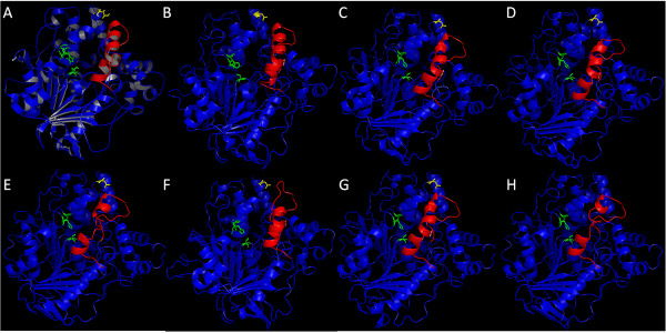
Figure 2.

Tridimensional structures/models of the proteins in its open conformation. (A) Lip3 from C. rugosa. Models, based in C. rugosa Lip3 structure, from (B) OPE and the six selected putative proteins (C) Altbr1, (D) Aspni5, (E) Necha2, (F) Neudi1, (G) Pyrtr1 and (H) Trire2. Lid region is indicated in red, the three catalytic residues are highlighted in green and the residue for a putative glycosylation site is marked in yellow.
