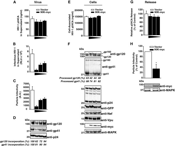
Figure 1.
Heterologous expression of 90K reduces the particle infectivity of HIV-1 progeny by interfering with the maturation and incorporation of the viral glycoproteins gp120 and gp41. (A) 293T cells were cotransfected with 1.3 μg pBR.HIV-1 IRES GFP and various amounts of pcDNA6.90K-myc (1.3, 0.4, 0.15 μg) or empty vector. The total amount of DNA was kept constant (2.6 μg). Supernatants were analyzed for amounts of released HIV-1 capsid antigen by anti-p24 capsid ELISA and (B) infectious HIV-1 by applying a TZM-bl luciferase assay. (C) Particle infectivity was defined as infectivity per ng released capsid. Shown are arithmetic means ± S.D. of triplicates. (D) Sucrose cushion-purified virions were analyzed by immunoblotting. Percentages indicate the relative gp120 and gp41 incorporation, as measured by Infrared imaging-based quantification of the amount of gp120 and gp41 per p24, respectively. The values of incorporation in absence of 90K expression were set to 100%. (E) Corresponding cell lysates were analyzed for cell-associated p24 capsid by ELISA. (F) Cell lysates were analyzed by immunoblotting using the indicated antibodies. Numbers indicate the efficiency of gp120 and gp41 processing, respectively, calculated as the signal ratio of gp120 relative to (gp120 + gp160), or of gp41 relative to (gp41 + gp160), respectively. (G) Capsid release was calculated as p24 capsid antigen in the supernatant (p24Sup) to total p24 capsid (p24Cell + p24Sup). The condition without 90K was set to 100%. (H) SupT1 cells were electroporated with pBR.HIV-1 IRES GFP (10 μg) and pcDNA6.90K-myc or empty vector (10 μg). Three days post transfection, supernatants were harvested and analyzed for particle infectivity by applying a TZM-bl infectivity assay and an anti-p24 capsid ELISA of the sucrose cushion-purified supernatants. Western Blot analysis of the producer cells was performed using the indicated antibodies.

