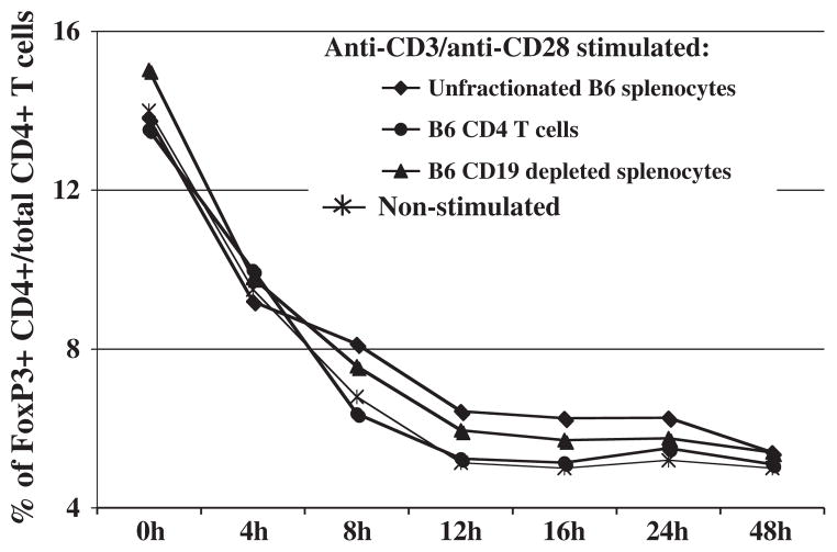
Fig. 2.
Decline of the percentages of CD4+ FoxP3+ Treg cells upon in vitro incubation. Either unfractionated splenocytes, positively-selected CD4+ T cells, or negatively-selected CD4+ T cells (CD19-depleted splenocytes) were cultured in parallel with in vitro stimulation. Cells were harvested at the indicated times post in vitro stimulation with plate-bound anti-CD3 and soluble anti-CD28 for anti-CD4 and anti-CD154 staining. FoxP3+ cells were identified using intracellular FoxP3 staining (see Materials and methods). Percentages of CD4+ FoxP3+ Treg cells are representative of three independent experiments.

