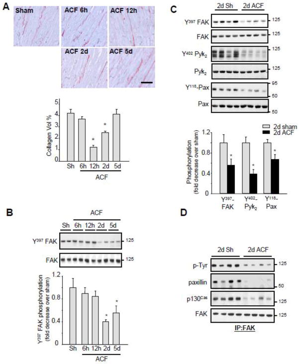
Figure 1. Acute ACF induces FA signaling downregulation.
(A) Top, Representative picrosirius red staining.(Bar = 40 μM) Bottom, Interstitial collagen accumulation as determined by morphometric analysis. (B–C) LV extracts from control and ACF operated rats were assessed for immunoblot analysis. Top, representative immunoblots (with each lane from a single gel exposed for the same duration). Bottom, fold induction, n=6 each group, *P<0.05 vs. sham. (D) LV lysates from sham or ACF animals were immunoprecipitated (IP) with anti-FAK antibodies and immunoblotted with anti-phosphotyrosine, p130Cas, paxillin or FAK antibodies.

