Figure 1.

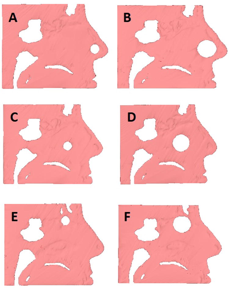
Cross-sectional sagittal images showing sizes and locations of the modeled perforations. 1 cm (A) and 2 cm (B) anterior perforations, 1 cm (C) and 2 cm (D) posterior perforations, and 1 cm (E) and 2 cm (F) superior perforations.

Cross-sectional sagittal images showing sizes and locations of the modeled perforations. 1 cm (A) and 2 cm (B) anterior perforations, 1 cm (C) and 2 cm (D) posterior perforations, and 1 cm (E) and 2 cm (F) superior perforations.