Figure 5.
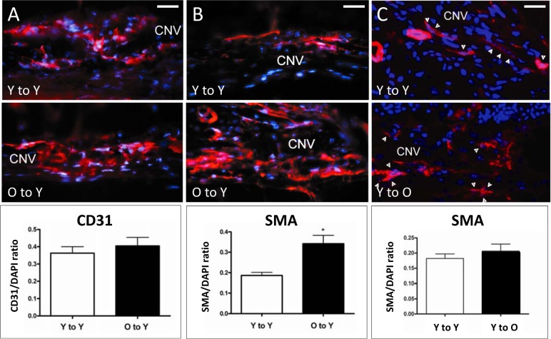
Immunofluorescence detection of endothelial and perivascular mesenchymal cells present in CNV lesions after BMT. Endothelial cells were stained with CD31 (left) and perivascular mesenchymal cells with SMA (center and right). In spite of the difference in lesion size, the frequency of endothelial cells (A) was not significantly different between the young-to-young (36.3 ± 0.04%) and the old-to-young group (40.5 ± 0.05%). In contrast, a significant increase (asterisk) in the frequency of SMA-expressing cells (B) was observed when the old-to-young group (34.2 ± 0.04%) was compared to the young-to-young group (18.7 ± 0.01%, t-test: P < 0.002). (C) No significant increase in the frequency of SMA-expressing cells (arrowheads) was observed between the young-to-young (18.22 ± 0.01%) and the young-to-old group (20.7 ± 0.02%, t-test: P = 0.360). Magnification: ×400; scale bars: 100 μm; CD31 (left) or SMA (center and right), red; DAPI, blue.

