
Figure 6.
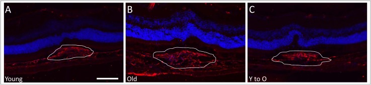
Levels of collagen type IV deposition present in CNV lesions. Cryosections with representative CNV lesions were stained with an antibody to collagen type IV to determine extent of fibrosis in the different groups. The amount of collagen IV deposition was greater in CNV lesions of old (B) compared to those of young (A) unmanipulated mice. In contrast, a decrease in collagen deposition was observed in CNV lesions in young-to-old mice (C) when compared to old unmanipulated mice. White lines outline neovascular lesions. Magnification: ×200; scale bar: 100 μm. Collagen type IV (red), DAPI (blue).