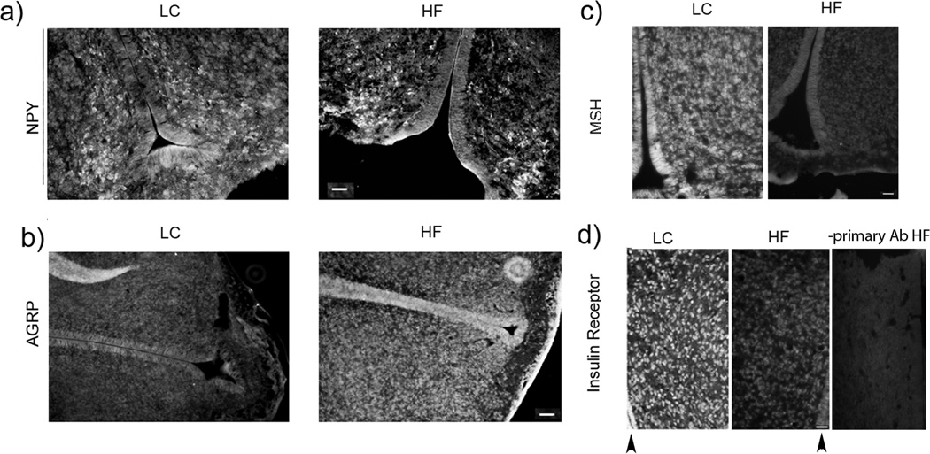
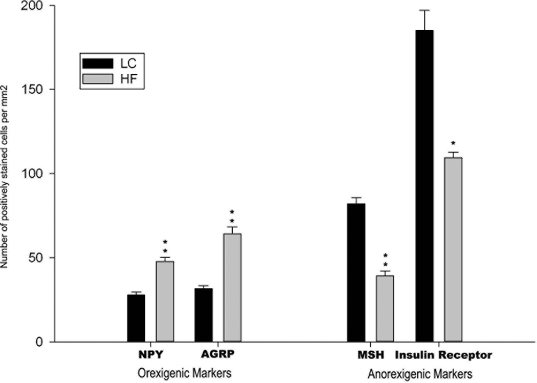
Fig. 1.
Histological analysis of the diencephalic region of term fetal MF and HF rats brain sections. Immunostaining of (a) NPY, (b) AGRP, (c) α-MSH and (d) insulin receptor (arrowheads indicate 3rd ventricle). Bar size: a- and b - 100 µm; c and d- 50 µm. (e) bars show average numbers of immunostained cells/mm2 counted on 4 brain sections from the corresponding brain areas. Difference between HF and MF sections were evaluated using Students’ t test: *P < 0.05; ** P<0.001.


