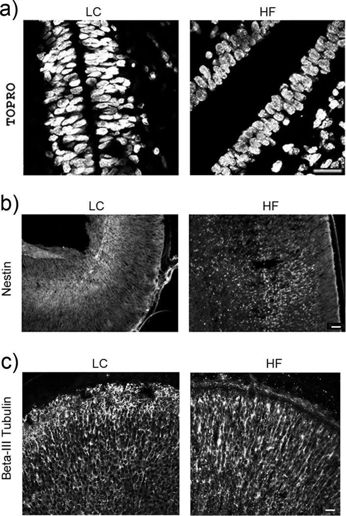
Fig. 2.
Histological analysis of the area of the 3rd ventricle and brain cortex. Immunostaining of (a) TOPRO stained cell nuclei in the area of the 3rd ventricle (neurogenic area), (b) Nestin-expressing neural progenitor cells in brain cortex and (c) β-III Tubulin-expressing immature neurons in brain cortex. Bar size: a - 50 µm; b and c - 100 µm.

