
Fig 2.
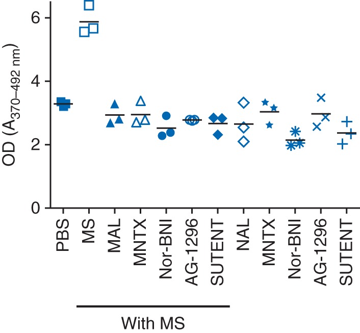
Mesangial cell proliferation shown as optical density (OD) in response to morphine and PDGFR inhibitors or OP antagonists. Mesangial cells were incubated for 48 h with 1 μM morphine in the presence or absence of 1 μM naloxone, 0.1 μM MNTX, 1 μM nor-BNI, 10 μM AG1296, and 2 μM sunitinib. Mean (sd), n=3.