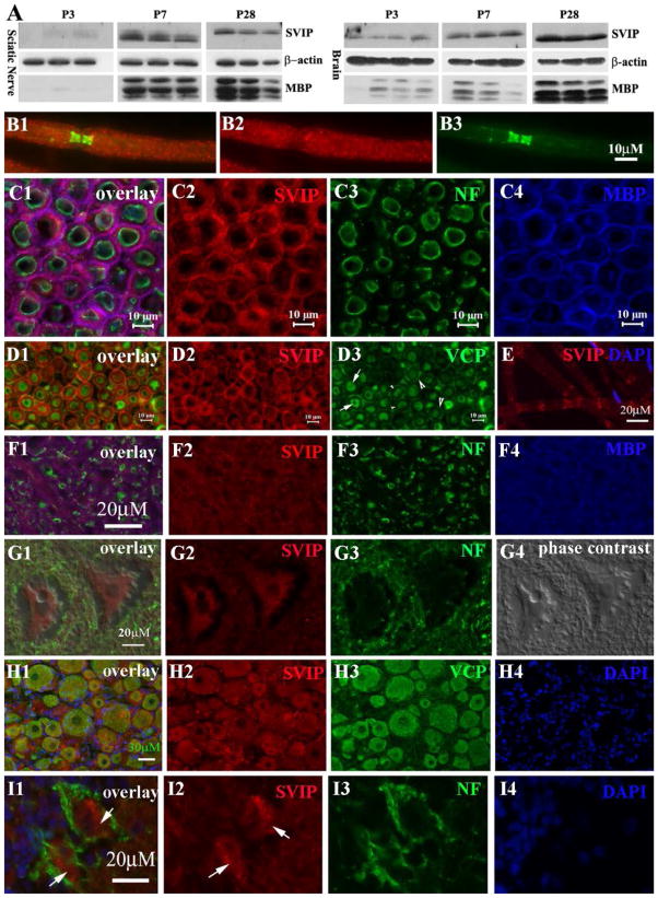
FIGURE 1. Expression of SVIP in the nervous system.
(A) Mouse tissues were taken at postnatal day 3, 7 and 28 and processed for Western blot analysis. SVIP was barely detectable at P3 and strongly increased at P7 and P28. A similar change was also observed for MBP. (B1–3) Adult rat sciatic nerves were teased into individual fibers and stained with antibodies against SVIP and Caspr, a paranodal marker. SVIP was localized on internodal myelin and weakly in paranodal myelin. (C1–4) SVIP was stained on the cryostat transverse sections of adult rat ventral and dorsal roots. Myelin was labeled by antibodies against MBP (blue color in C4) and axon was labeled by antibodies against phosphorylated neurofilaments (NF; green color in C3). SVIP overlapped well with MBP (pink color in C1), but not neurofilament, suggesting its localization in compact myelin. (D1–3) Cryostat transverse sections of adult rat sciatic nerves were stained with antibodies against SVIP and VCP. VCP was mainly expressed in axons (arrows in D3) and in the abaxonal cytoplasmic space of Schwann cell (large arrowheads in D3), but not in compact myelin (small arrowheads in D3). VCP does not overlap with SVIP in myelin (D1). E. Teased nerve fibers were stained for SVIP. A small fraction of myelinated nerve fibers showed SVIP expression in Schmidt-Lanterman incisures. (F1–4) Cryostat sections of adult rat spinal cord were stained with antibodies against SVIP and MBP. This image was taken from the ventral column (white matter) of the spinal cord. SVIP immunoreactivity (red color in F2) also overlapped with MBP (pink color in F1), but not with axons labeled by neurofilament (green color in F1). (G1–4) SVIP immunoreactivity was also found in the cytoplasm of spinal motor neurons (G2). (H1–4) Immunofluorescence staining was also performed on the cryostat sections of the adult rat dorsal root ganglion. Again, SVIP and VCP were well overlapped in the cytoplasm of neurons. (I1–4) Cryostat sections from the adult rat cerebellum were stained and showed SVIP expression in the cytoplasm of Purkinje cells (arrows in I1–2).

