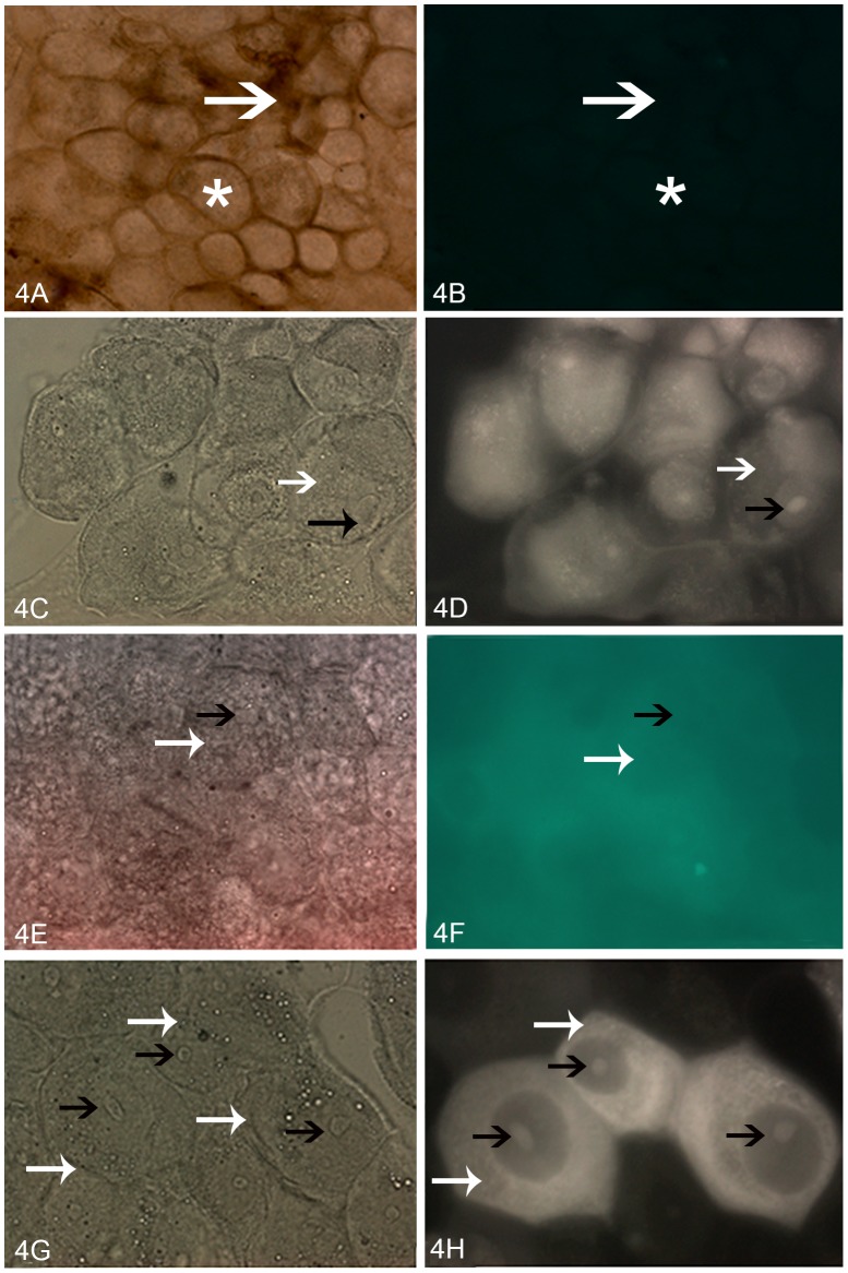
Figure 4. Photomicrographs of posterior midgut epithelial cells of fifth-instar R. prolixus incubated with biotin-labeled peptides.
(A) Light microscopy showing single-globe columnar epithelial cells(white star) and PMM (white arrow). (B) Fluorescence microscopy showing that no demarcation was observed after incubation with avidin-FITC-labeled conjugate alone. Light and fluorescence microscopy, respectively, of samples incubated with biotin-labeled TcSMUG L (C and D), biotin-labeled TcSMUG S (E and F), and biotin-labeled TSSA (G and H). Fluorescence of the surface and nucleolus of the midgut cells is indicated by white and black arrows (respectively). 400×.

