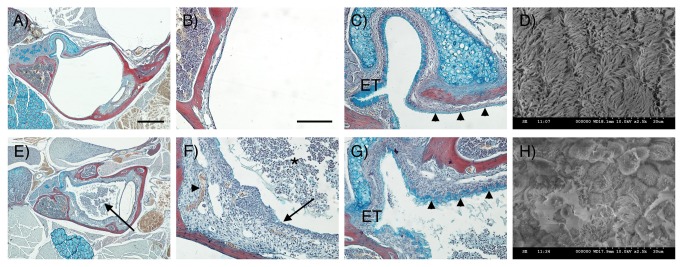
Figure 3. Morphological changes in MEC mucosa and epithelium in Df1/+ mice.
(A-C, E-G) Frontal trichrome-stained sections from 11.5-week-old mice showing the middle ear cavity. (D, H) SEM images of middle ear epithelium. (A-D) WT. (E-H) Df1/+ with signs of OM. (A) The middle ear cavity is air-filled in the WT. (B) The mucosa is a thin layer lining the auditory bulla. (C) At the entrance of the Eustachian tube (ET) high levels of alcian blue staining are observed indicating mucin production. Further into the middle ear away from the orifice, staining is less distinct in the WT (arrowheads). (D) A thick lawn of cilia is observed overlying the epithelium near the ET orifice. (E) In Df1/+ mice the middle ear cavity is filled with effusion (arrow). (F) Df1/+ mice show signs of inflammation such as effusion with infiltrated inflammatory cells (asterix), a thickened mucosa (arrow) and hypervascularisation (arrowhead). (G) In addition, increased alcian blue staining is observed within the middle ear at a distance from the ET indicating increased mucin production in Df1/+ mice (compare C and G, arrowheads). (H) Df1/+ mice with OM show reduced numbers of cilia that appear shortened and rarefied. Dorsal is top in A-C, E-G. Scale bar: 500 μm (A, E), 100 μm (B, C, F, G).

