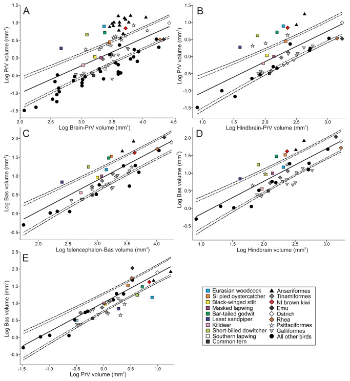
Figure 5. PrV and Bas volumes relative to the volume of the brain and hindbrain.
Scatterplots of the log-volume of the principal sensory trigeminal nucleus (PrV) and the nucleus basorostralis (Bas, all measurements in mm3). Data from [8] and [37] is included in the analysis. PrV is regressed against A: Brain-PrV volume and B: hindbrain-PrV volume and Bas against C: telencephalon-Bas volume and D: hindbrain volume. E shows Bas regressed against PrV. The solid lines indicate the least-squares linear regression lines and the dotted and broken lines indicate the phylogeny-corrected 90 and 95% confidence interval, respectively. Symbols and colors represent birds and orders as shown in the legend.

