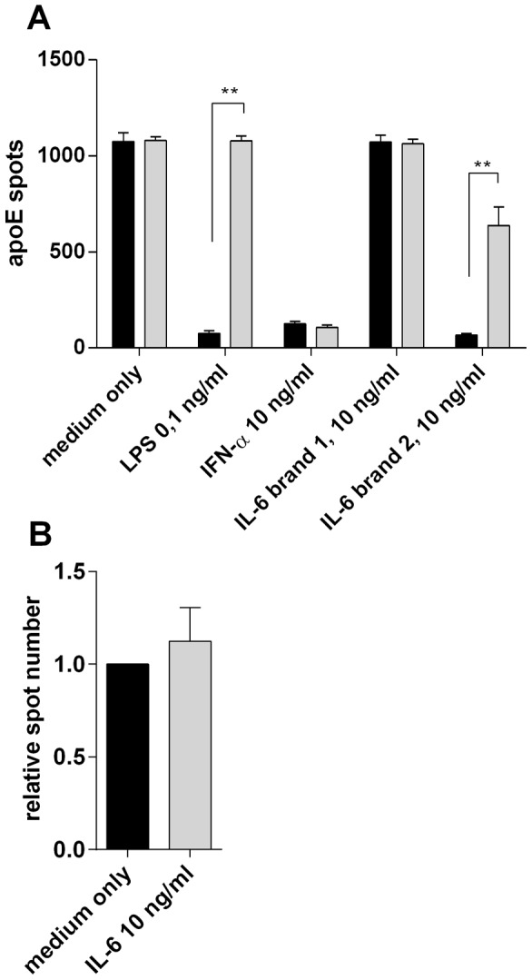
Figure 3. Verified lack of IL-6 effect on apoE production in the presence of the LPS-inhibitor Polymyxin B.

A) Example of how contaminating LPS in cytokine preparations can affect apoE production. TGF-β-treated PBMC (100×103 cells/well) were incubated for 20 hours in the presence of LPS (0.1 ng/ml), IFN-α, or either of two preparations of IL-6 (brand 1 and 2). All cytokines were used at 10 ng/ml. Cells were incubated with (grey bars) or without (black bars) Polymyxin B (10 µg/ml). Results shown are from one representative donor (means ± SD of triplicate). (B) PBMC (100×103 cells/well) were incubated for 20 hours in the presence of TGF-β (10 ng/ml) and Polymyxin B (10 µg/ml) ( = medium only) with or without IL-6 (10 ng/ml). Relative spot numbers for each donor were calculated by dividing apoE spot numbers of IL-6 treated PBMC with the number of spots obtained without IL-6. Results shown are means ± SD of 4 donors.
