
Figure 5. Inhibition of ApoE secretion by PBMC in the presence of activated T cells.
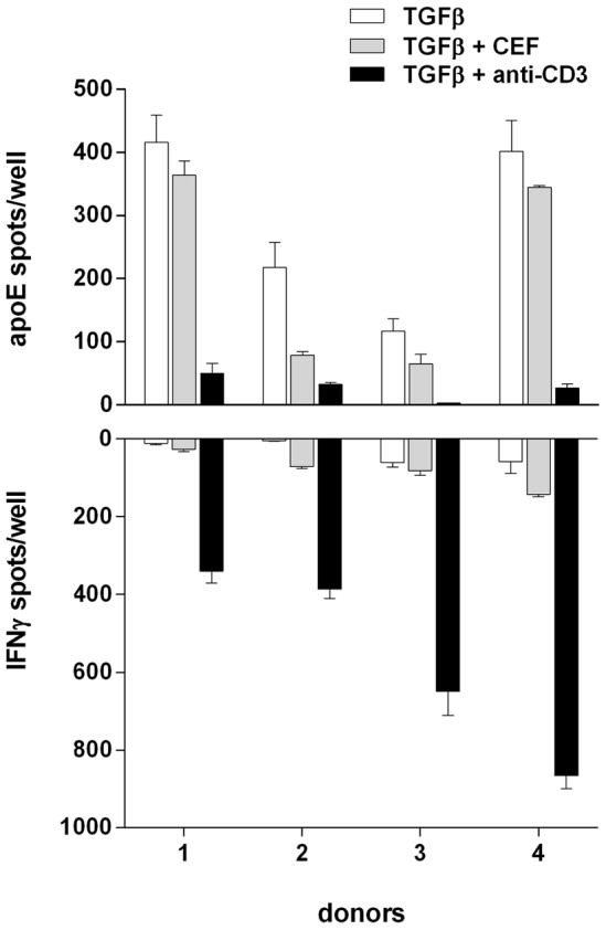
PBMC (100×103 cells/well) from 4 different donors were incubated for 20 hours in the presence of TGF-β (10 ng/ml) and Polymyxin B (10 µg/ml) with or without anti-CD3 (100 ng/ml) or CEF (2 µg/ml) and analysed for secretion of apoE (top) and IFN-γ (bottom) in the ELISpot assay. Values represent means ± SD of triplicates.