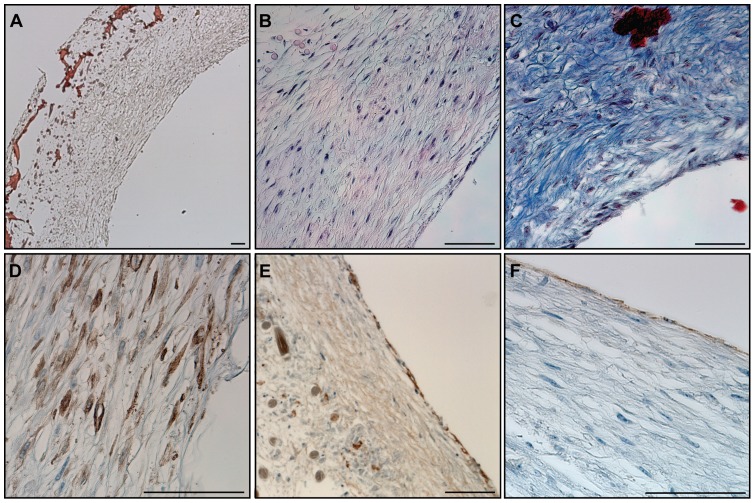
Figure 2. Histological structure of an engineered artery equivalent.
A) Haematoxylin-Sudan staining demonstrated the formation of tissue in and on the surface of a PGA/P4HB scaffold as well as the scaffold’s partial degradation. B) H&E staining revealed dense tissue formation composed of cells and extracellular matrix. C) The secretion of collagen was observed after Masson’s trichrome staining. The expression of α-smooth muscle actin (α-SMA) (D) confirmed the smooth muscle phenotype of the cells in the inner layer. Collagen IV positive staining (E) demonstrated the secretion of basement membrane and CD31 positive staining (F) confirmed the presence of an endothelial cell monolayer on the luminal side of the bioengineered artery equivalent. Bars represent 100 µm.

