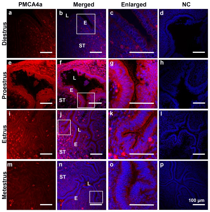
Figure 3. Indirect Immunofluorescence of PMCA4a in the murine endometrium of the uterus during the estrous cycle.
Using frozen sections PMCA4a immunoreactivity (red) was detected in the uterine luminal epithelium in pro-estrus (e - g) and estrus phases (i - k) but not during metestrus (m - o) and diestrus (a - c). The nuclei were visualized by staining with Draq-5 (blue). Negative controls (NC) in PBS or IgG are shown in d, h, l, and p. The images were captured using confocal microscopy and a 20x (a plan-Apochromatic) objective lens. LE = luminal epithelium; L= lumen; ST= stroma. Bar = 100 µm (same scale for all micrographs, and 200 µm for insets).

