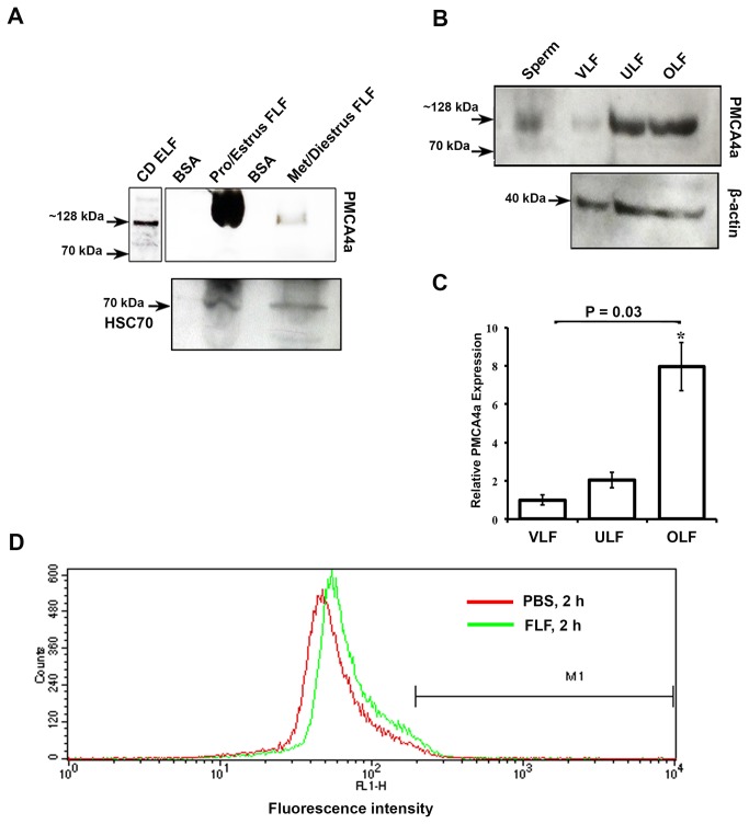
Figure 5. Detection of PMCA4a in reproductive luminal fluids and its acquisition on caudal sperm.
A) Representative Western blot of FLFs collected during pro-estrus and estrus and metestrus and diestrus (40 µg proteins loaded). The ~128 kDa PMCA4a is seen in pro-estrus and estrus and is marginally present at metestrus and diestrus. Caudal epididymal luminal fluid was used as a positive control. The membrane was stripped and re-probed for HSC70 as a loading control. B) Western blots of VLF, ULF, and OLF recovered after superovulation demonstrate the presence of the ~128 kDa PMCA4a. Sperm protein was used as a positive control. The membrane was stripped and re-probed for β-actin as a loading control. C) Quantitation of Western blot data shown in B; the relative expression was determined using VLF as 1. The data represent the mean (±SEM) of a minimum of three independent experiments, and the intensity was quantified by Image J software. ANOVA and t-tests were performed on the mean and P values were calculated. *P = 0.03 indicates a significantly increase amount of PMCA4a in OLF compared to that in VLF. D) A peak shift of fluorescence intensity to the right, indicates increase amounts of PMCA4a in sperm incubated in FLF compared to PBS for 2 h and treated as described in Materials and Methods.

