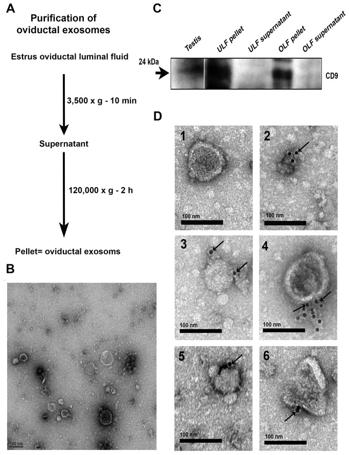
Figure 6. Characterization of membranous vesicles in OLF.
A) Protocol used to isolate oviductal exosomes by ultracentrifugation of oviductal fluids. B) TEM of negative staining for the particulate fraction from OLF reveals the presence of membranous vesicles ranging in size from 25-100 nm in diameter. C) Western blots detected CD9 (24 kDa) in protein extracts from membranous vesicles removed from OLF and uterosomes, but not in the supernatants. Testis protein was used as a positive control. Each lane contains 40 µg of protein. Results are representative of three different experiments. D) Immunogold labeling (6 nm gold particles) of CD9 is shown in oviductal membranous vesicles termed “oviductosomes”. Gold particles on individual oviductosomes are seen arrowed in 2-6 on the exterior of the membrane. In the absence of primary antibodies and the presence of rat IgG, gold particles were absent (1), indicating the specificity of the antibody. Scale bar =100 nm in panel B, D.

