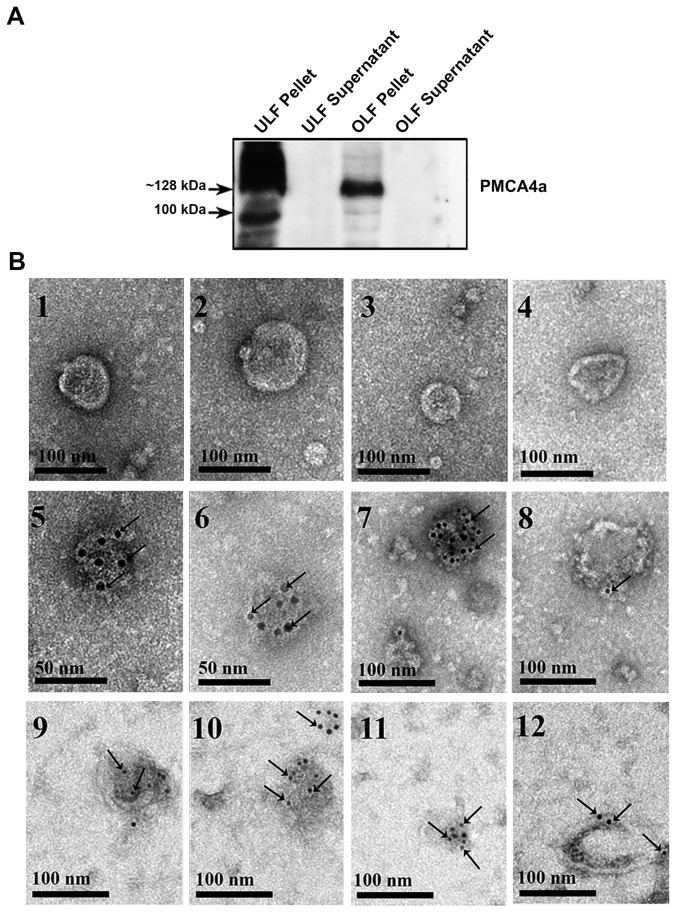
Figure 7. Immunodetection of PMCA4a in oviductosomes and uterosomes.
A) Nitrocellulose membrane with CD-positive pellets and supernatants (Figure 6C), when stripped and re-probed with PMCA4a antibodies in a Western blot, revealed the presence of the ~128 kDa PMCA4a band in the pellets only. A band of unknown origin at ~100 kDa is also seen in the uterosomes. Each lane contains 40 µg of proteins. Results are representative of three experiments. B) Immunogold labeling (6 nm gold particles) of PMCA4a is shown in oviductosomes in 5-8 and in uterosomes in 9-12. Gold particles localized on the cytoplasmic-side of the membrane are seen arrowed. They were rarely seen elsewhere on the grids. In the absence of primary antibodies and the presence of rabbit IgG, gold particles were absent (1-4) indicating the specificity of the antibody. Scale bar= 50-100 nm.

