
Figure 8. PMCA4a uptake by caudal sperm via incubation in exosomes reconstituted in PBS.
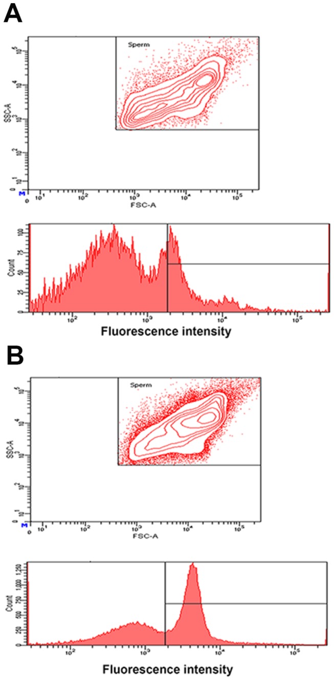
Exosomes were isolated from FLFs after superovulation. A) Flow cytometric analysis of sperm co-incubated in PBS (negative control). The contour plot above reveals two subpopulations of sperm, as represented by the small inner circles at top right and bottom left. The graph shows that the majority of the fluorescence falls below the gated region. B) Sperm incubated in FLF exosomes (2 mg/ml protein) show a unimodal distribution (a single inner circle) and a ~3 fold increase of fluorescence intensity (peak shift to the right) compared to A in the gated region. The co-incubation period was 3 h for both A and B.