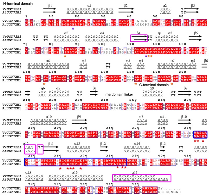
Figure 1. Structure-based sequence alignment of VvUGT72B1 and AtUGT72B1.

Secondary structure elements are shown above the alignment. Conserved residues are highlighted. The UGT signature PSPG motifs are enclosed in a bold blue box. Conserved residues involved in sugar donor binding are indicated with a red asterisk (*) below the alignment. Conserved residues interacting with the acceptor are indicated with a purple asterisk. Residues involved in enclosing TCP in AtUGT72B1 are indicated with a saffron yellow asterisk. “α” represents an α-helix, “β” represents a β-sheet, “TT” represents a β-turn, and “η” represents 3.10 helix. This figure was produced using ENDscript [31].
