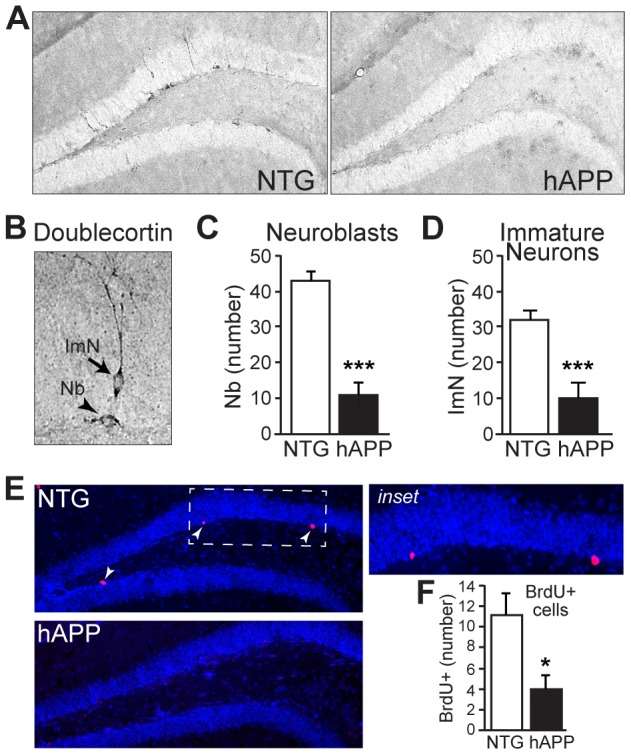
Figure 4. 13–15 month old hAPP mice exhibit decreased neurogenesis.

A, Micrographs of doublecortin immunostaining in coronal brain sections from NTG and hAPP mice. B, High magnification micrograph illustrating doublecortin-positive neuroblasts (Nb) and immature neurons (ImN). C–D, Quantification of doublecortin expression demonstrates significant decreases in neuroblasts (C) and immature neurons (D) in hAPP mice relative to NTG controls. E–F, BrdU labeling of dividing cells in the subgranular zone demonstrates fewer dividing cells in the subgranular zone of hAPP mice. Arrowheads, BrdU-labeled cells. n = 11–12/genotype. *p<0.05, ***p<0.001.
