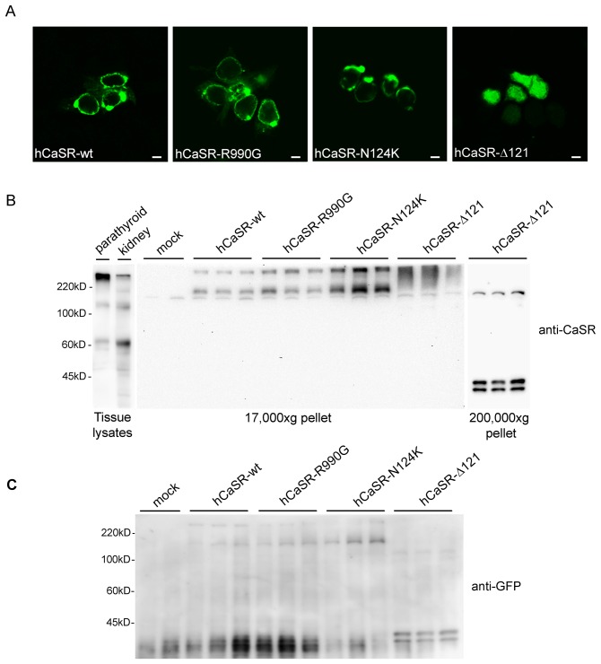
Figure 1. Expression and localization of Calcium Sensing Receptors (CaSR) in HEK-293 cells.
(A) Confocal microscopy showing cellular localization of hCaSR-wt and the gain-of-function variants hCaSR-R990G and hCaSR-N124K. hCaSR-wt as well as its active variants, were mainly expressed at the plasma membrane. In contrast, hCaSR-Δ121, corresponding to a loss-of-function truncated protein, localized intracellularly. The scale bar corresponds to 5 µm. (B) Western blotting analysis. Equal amounts of proteins (60 µg) isolated from tissue lysates (parathyroid and rat kidney) or from cell 17,000×g pellet were immunoblotted and probed with monoclonal antibodies against CaSR (N-terminal, 1∶800). In lysates isolated from rat parathyroid and rat kidneys a band at approximately 135 kDa and an aggregate at higher molecular weight was stained. An immunoreactive band of ≈170 kDa, a band greater than 220 kDa marker and likely corresponding to higher molecular mass forms of the receptor were detected only in cells transfected with hCaSR-wt, hCaSR-R990G and hCaSR-N124K. The higher molecular weight observed with respect to native tissues is due to the presence of the GFP tag. In cells transfected with hCaSR-Δ121, the antibody stained bands at approximately 40 kDa in a 200,000×g pellet consistent with a prominent intracellular distribution. (C) Cell extracts from cells transfected with hCaSR-wt, hCaSR-R990G, hCaSR-N124K and hCaSR-Δ121 were immunoblotted and probed with a monoclonal anti-GFP antibody (1∶5000). Anti-GFP antibody stained bands at the same molecular weight as those revealed by anti CaSR antibody confirming the specificity of the revealed bands.

