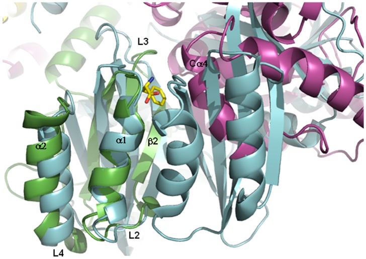
Figure 10. Superimposition of the Phe-bound prephenate dehydratase (PDT) and the tetrameric model of the hPAH.
The Phe-bound PDT from Chlorobium tepidum TLS (PDB code 2QMX) is drawn in cyan. The RD of a subunit of hPAH is drawn in green, whereas the catalytic domain of the adjacent subunit is drawn in violet. The bound Phe ligand is shown as a yellow stick.

