
Figure 2. Localization of active Ras in glucose-growing cyr1Δ pde2Δ yak1Δ cells, before and after addition of cAMP.
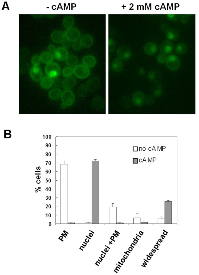
(A) cyr1Δ pde2Δ yak1Δ cells transformed with YEpeGFP-RBD3 were grown in medium containing 2% glucose at 30°C until exponential phase and then photographed with a Nikon fluorescence microscope, before and 45 min after addition of 2 mM cAMP. (B) Subcellular distribution of eGFP fluorescence.