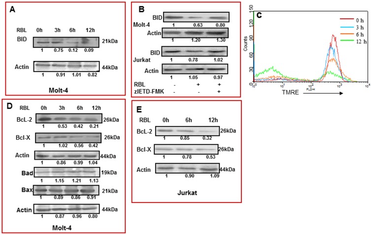
Figure 7. Cleavage of BID, loss of MMP, change in expression of anti- and pro-apoptotic proteins.
(A) Molt-4 cells were treated with RBL (5 µg/ml) for different time periods and the effect on 21kDa uncleaved Bid was studied by western blot analysis. Actin was used as loading control. The fold differences were compared to 0 h control. (B) Molt-4 and Jurkat cells were treated with RBL(5 µg/ml) for 12 h in the presence or absence of caspase-8 inhibitor (zIETD-FMK) and truncation of BID was assessed using western blot analysis. (C) Molt-4 cells treated with RBL for different time periods –0 h (red line), 3 h (blue line), 6 h (orange line), 12 h (green line), were incubated with TMRE dye for 20 min prior to harvesting and flow cytometry analysis was performed. Loss in mitochondrial membrane potential was recorded as decrease in the red fluorescence (MFI) on FL-2 channel. The overlay is representative of three similar experiments. (D) Molt-4 cells were treated with RBL (5 µg/ml) for different time durations and the expression of Bcl-2, Bcl-X, Bad and Bax were determined by western blot analysis. (E) Jurkat cells were exposed to RBL (5 µg/ml) for different time durations and the expression of Bcl-2 and Bcl-X was assessed. Actin was used as loading control. The fold differences were compared to 0 h control.

