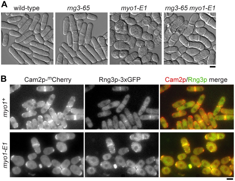
Figure 7. Destabilizing Myo1p motors leads to recruitment of Rng3p to patch structures.
The -E1 mutation was introduced into myo1 (G308R) in the genome to generate the myo1-E1 strain. A) Representative DIC images of wild-type, rng3-65, myo1-E1, and rng3-65 myo1-E1 cells following growth on YE5S media at 25°C. The double mutant was viable and exhibited morphology defects that were a combination of those observed in each of the single mutants (i.e. cells were elongated like rng3-65 cells and somewhat swollen like myo1-E1 cells). B) Myo1p (via its light chain Cam2p) and Rng3p localization in wild-type myo1+ (top) and myo1-E1 mutant (bottom) cells grown in YE5S media at 25°C. Left: single plane epi-fluorescence images of Cam2p-mCherry, center: single plane epi-fluorescence images of Rng3p-3xGFP, and right: merged images of Cam2p (red) and Rng3p (green) signals. Bars: 4 µm.

