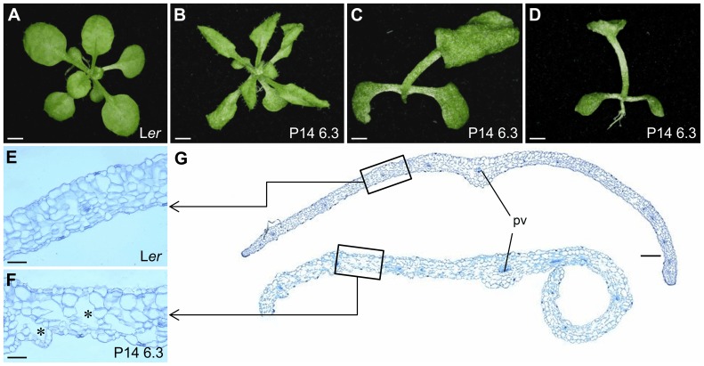
Figure 1. Leaf morphology and histology in the P14 6.3 line.
(A, B) Rosettes of (A) Ler and (B) P14 6.3 plants. (C) Top and (D) lateral views of a P14 6.3 plant showing fusion of the first pair of vegetative leaves. (E, F) Transverse sections of the central region of the lamina, midway between the leaf margin and the primary vein of (E) Ler and (F) P14 6.3 third-node leaves. Asterisks indicate large intercellular air spaces. (G) Margin to margin transverse sections of third-node leaves. pv: primary vein. Pictures were taken (A–D) 22 das and (E–G) 21 das. Scale bars: (A, B, D) 2 mm, (C) 1 mm, (E, F) 50 µm and (G) 200 µm.

