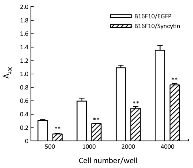
Figure 4.

Comparison of cell proliferation between B16F10 cells stably transfected with pEGFP-N1 (B16F10/EGFP) and pEGFP/syncytin (B16F10/syncytin). Indicated numbers of cells were seeded in 96-well plates and further cultured for 48 h. MTS assay was used to detect the proliferation rates of the cells. **, P<0.01 using Student’s t-test.
