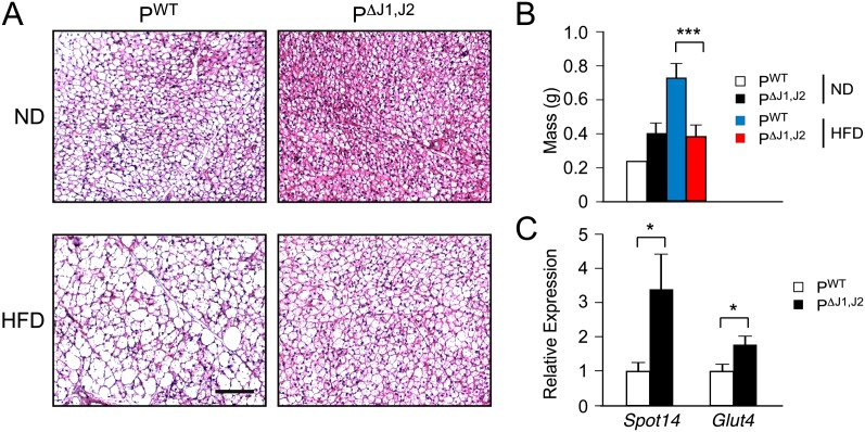
Figure 2.
JNK in the anterior pituitary gland promotes HFD-induced hypertrophy of brown adipose tissue. (A) PWT and PΔJ1,J2 mice were fed an ND or a HFD (16 wk). Sections of interscapular brown adipose tissue were stained with hematoxylin and eosin. Bar, 125 μm. (B) Total interscapular brown adipose tissue mass was measured (mean ± SEM; n = 14∼20). (***) P < 0.001. (C) The expression of Spot14 and Glut4 mRNA in brown adipose tissue was measured by quantitative RT–PCR assays (mean ± SEM; n = 10∼12). (*) P < 0.05.

