Figure 3.
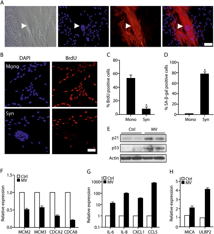
MV induces cell fusion and senescence in normal human fibroblasts. (A) The MV-infected IMR-90 cells were stained with anti-SSPE antibody that recognizes MV proteins (red) and counterstained with DAPI. BrdU incorporation (B,C) and SA-β-gal activity (D) were assessed in IMR-90 cells infected with MV in syncytium (Syn) and mononuclear cells (Mono). (E) Protein content of p53 and p21 was assessed by immunoblotting in MV-infected or mock-infected (Ctrl) IMR-90 cells. Expression of MCM2, MCM3, CDCA2, and CDCA8 (F); IL-6, IL-8, CXCL-1, and CCL5 (G); and MICA and ULBP2 (H) in MV-infected or mock-infected (Ctrl) IMR-90 cells assayed by quantitative RT–PCR. Values are means + SEM; (*) P < 0.05.

