Figure 1.
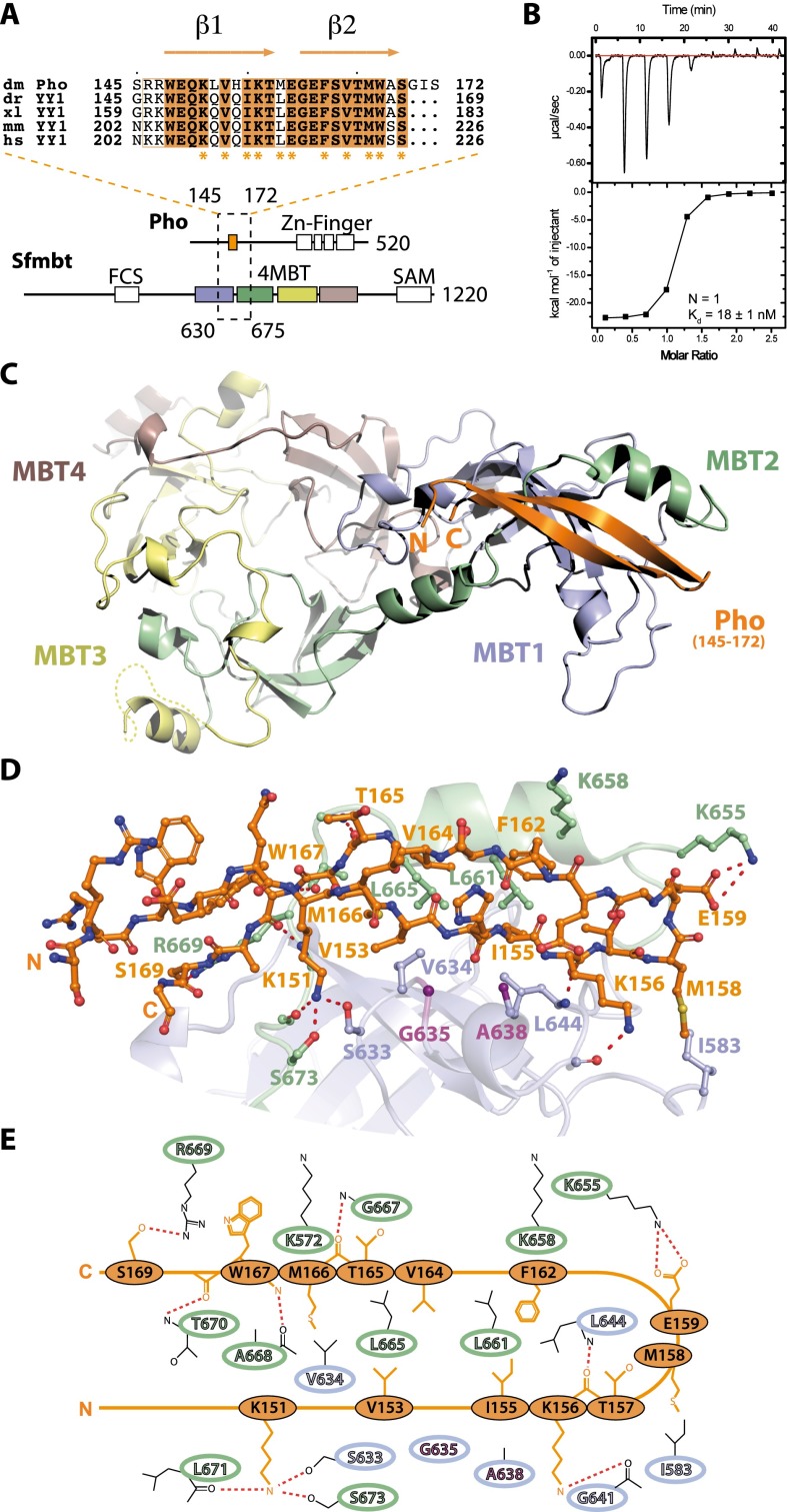
Biophysical and structural characterization of the Pho spacer:Sfmbt 4MBT interaction. (A, top) Sequence alignment of the Drosophila melanogaster Pho spacer region (dm, Q8ST83, orange) with the YY1 orthologs from Danio rerio (dr, Q7T1S3), Xenopus laevis (xl, Q6DDI1), mice (mm, Q00899), and humans (hs, P25490). Residues involved in the interaction with the Sfmbt 4MBT domain are indicated with asterisks. (Bottom) Pho and Sfmbt domain architecture. Pho spacer:Sfmbt 4MBT-interacting regions are enclosed by a dashed rectangle, and the first and last residue of the interacting regions are given. Sfmbt MBT repeats 1–4 are colored. (B) ITC data of the Pho spacer:Sfmbt 4MBT interaction. (C) Overview of the miniPhoRC complex crystal structure as a ribbon diagram presentation. (D) Close-up view of the Pho spacer:Sfmbt 4MBT interaction. Interacting residues of the Pho spacer and the Sfmbt 4MBT domain are depicted. Gly635 and Ala638 in the Sfmbt clamping helix are highlighted (purple). (E) Schematic representation of the Pho spacer:4MBT domain interaction.

