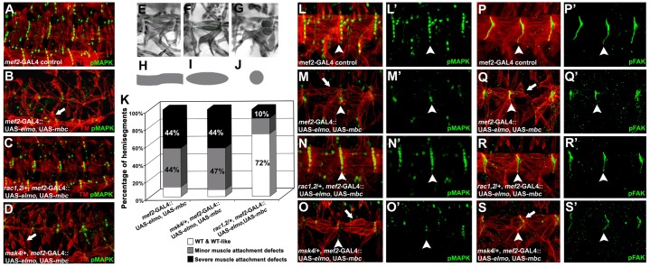
Fig. 4.
The muscle-specific Elmo–Mbc→Rac signaling module non-autonomously signals to maintain tendon cell identity through pMAPK. (A–D) Representative embryos detailing the lateral musculature (TM; red) and tendon cell nuclei (pMAPK; green) in stage 16–17 embryos. Muscle-specific coexpression of Mbc and Elmo result in severe muscle attachment defects with smaller MASs (B; arrows) than mef2-GAL4/+ control embryos (A). Removal of one copy of endogenous rac1J1rac2Δ(C), but not msk4 (D), restores normal muscle attachment. (E–K) For quantification (K), the muscles in hemisegments A1–4 in each embryo (n = 200) were scored as WT (E,H); minor (F,I); or severe muscle detachment (G,J). (L–S′) High magnification of two hemisegments of the embryonic ventral musculature (TM; red) co-stained with the muscle or tendon cell markers, pFAK and pMAPK, respectively (green). In mef2-GAL4 controls, pFAK properly localizes to MASs (P,P′) with 6–7 pMAPK(+) tendon cells at the segmental border (L,L′). An excess of Elmo and Mbc in muscles results in smaller MASs, indicated by pFAK staining (Q,Q′) and a decrease in the number of pMAPK(+) cells (M,M′). The pFAK and pMAPK expression patterns are rescued to WT with the decrease of endogenous rac1 and rac2 (N,N′, R,R′), whereas they remain unaltered upon loss of one copy of msk (O,O′, S,S′).

