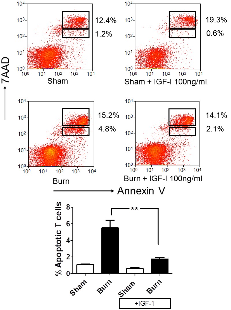
Figure 6.

IGF-I can act to prevent thymocyte apoptosis caused by burn injury. Wild-type female C57/BL6 mice received either a 20% TBSA or sham treatment (n=6/group). Thymocytes were harvested at 6 hours post burn from each mouse and placed thymocytes in culture in the absence or presence of 100 ng/ml IGF-I. Eighteen hours after burn, apoptosis was assessed by staining with anti-CD8 (APC), anti-CD3 (FITC), Annexin V conjugated to PE and 7-Aminoactinomycin D (7AAD) in PBS containing calcium. The degree of apoptosis was assessed by four color flow cytometry, quantifying the percentage of CD8+CD3+T cells that are apoptotic (defined by 7AAD-,lo Annexin V+ staining). Top panel, representative flow cytometry of each group. Bottom Panel, collated groups, plotting percentage of CD8+CD3+T cells that were apoptotic, where statistical significance is indicated by **, p<0.005 by two way ANOVA.
