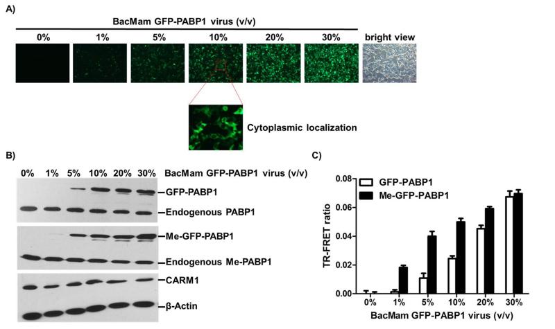
Figure 3. BacMam virus-mediated GFP-PABP1 protein can be methylated by CARM1 and TR-FRET signal can be measured in MCF7 cells.
(A) MCF7 cells were infected with the indicated amounts of BacMam GFP-PABP1 virus (4000 virus particles per microliter, indicated as volume/volume) in a 6-well plate for 24 hours. GFP imaging showed that BacMam virus-mediated GFP-PABP1 was expressed in a virus dose-dependent manner. (B) MCF7 cells were infected as in (A). Whole cell extracts were analyzed by western blots using antibodies against PABP1, Me-PABP1, CARM1 and β-actin. (C) MCF7 cells were infected as in (A) in a 384-well assay plate (7500 cells per well, 30 μl in total). TR-FRET measurement with Me-PABP1 or PABP1 primary antibody and Tb-2nd antibody indicated the increase of TR-FRET ratio as the dose of BacMam GFP-PABP1 virus increased. Background signal from Tb-2nd antibody alone was subtracted. The fold of Me-GFP-PABP1/ GFP-PABP1 activation is marked on the top of each condition. Data are mean ± SD of three independent experiments.

