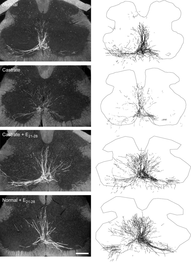
Figure 3.
(Left) Darkfield digital micrographs of transverse sections through the lumbar spinal cord of a normal male (top), an untreated castrate (second from top), a castrate treated with an estradiol implant from P21–P28 (Castrate + E21–28; second from bottom), and a normal male treated with an estradiol implant from P21–P28 (Normal + E21–28; bottom) after BHRP injection into the left BC muscle at P28. (Right) Computer-generated composites of BHRP-labeled somata and processes drawn at 320 µm intervals through the entire rostrocaudal extent of the SNB; these composites were selected as they are representative of their respective group average dendritic lengths. Scale bar = 250 µm.

