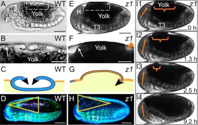
Fig. 6. The apical–basal orientation of EE tissue contraction affects DC geometry.
(A–D) WT, (E–I) Tc-zen1RNAi. (A,E) Whole embryo lateral views (transmitted light micrographs; fixed, A; live, E). (B,F) Enlargements of boxed regions in A,E. Unlike the folded serosa, the thin amnion (arrows) can hardly be distinguished from the underlying yolk. The arrowhead marks the Tc-zen1RNAi amniotic crease. For cell shape data, see Fig. 2B,D1. (C,G) Corresponding midsagittal schematics: serosal apical contraction produces a hollow structure, whereas basal Tc-zen1RNAi amniotic contraction inappropriately displaces yolk (color coding as in Fig. 1A, black for emphasis only here). (D,H) Subsequently, the overall geometry of the closing EE area is affected (triangle orientation; shown in dorsal–lateral aspect, reproduced from Fig. 1E,M). See supplementary material Table S1 for quantification. (I1–I4) Still images from supplementary material Movie 9: the yolk bulge (curly bracket) is eventually incorporated anteriorly, concomitant with an embryonic flexure that stretches the dorsum (note changing angle of the straight line between head landmarks). This postural change also occurs in WT (supplementary material Movie 1). Anterior is left and dorsal up in all panels. Abbreviations as in previous figures. Scale bars: 100 µm (A,D,E,H,I), 50 µm (B,F).

