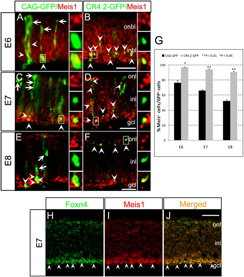
Fig. 6. Meis1 protein is present in CR4.2-GFP+ and Foxn4+ cells.
Chick retinas were electroporated with either the control CAG-GFP construct or CR4.2-GFP construct at E4. Transfected retinas were harvested at E6(A-B), E7(C-D), E8(E-F), sectioned, and immunostained for GFP (green) and Meis1 (red). (G) Quantification showed that a significantly high percentage of CR4.2GFP+ cells were co-labeled with Meis1as compared with the control CAG-GFP+ cellsat E6, E7 and E8 arrowheads in B, D, and F). Error bars represent standard error of the mean. Each histogram represents the mean ± s.d.; n≥3. (H-J) At E7, the majority of Foxn4+ cells were co-labeled with Meis1 staining (arrowheads). ONBL, outer neuroblastic layer; INBL, inner neuroblastic layer; ONL, outer nuclear layer; INL, inner nuclear layer; GCL, ganglion cell layer. Scale bars = 20 µm.

