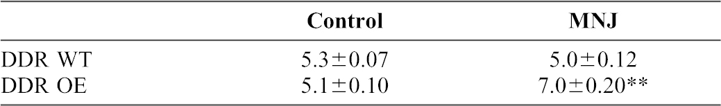
Table 2. Cell surface abundance of β1 integrin measured by flow cytometry.
DDR wild type and DDR over-expressing cells were treated with deoxymannojirimycin (MNJ) as indicated in the methods section. Cells were separated from plates with Versene and non-permeabilized, single cell suspensions were stained with KIM6 antibody that recognizes β1 integrin and counter-stained with FITC-conjugated goat anti-mouse antibody. Cells (10,000) were analyzed by flow cytometry and the mean±standard error of the mean fluorescence intensity are indicated. Significant differences of fluorescence intensities of β1 integrin staining between indicated groups are marked by ** for P<0.01.